Everything You Need to Know About Facetectomy Surgery
The facet joint, also known as the zygapophysial joint, is a movable joint located between the inferior and superior articular processes of the vertebra. Each vertebra contains two facet joints. These joints allow for spine mobility, and help a person bend forward, stand straight, and rotate left and right.
Facets joint degeneration occur due to age or through injury. The cartilage tissue around these joints erode or tear. When this happens, the bones rub together, restricting movement of the spinal column and compressing the spinal nerve roots housed in the spinal canal underlying the spinal column.
Spinal nerve root compression results in severe pain. The pain remains localized at first, but as the amount of pressure exerted on the spinal nerve roots by the bones increases, the pain spreads and soon causes loss of mobility.
Age related degeneration of facets joints is a natural process that cannot be reversed. It is, however, possible to ease the pressures on the spinal nerves, and consequently alleviate the pain through facetectomy, an invasive surgical procedure. Facetectomy surgery removes one or more facets to preserve nerve functioning.
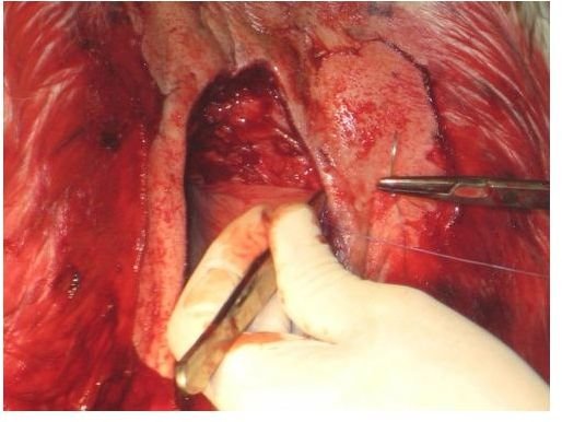
Image Credit: flickr.com/Vet Moves
Treatment
The first treatment to alleviate the pain caused by denigration of facet joints and compression of spinal nerve roots are physical therapy, heat therapy, and administration of pain relievers.
Facetectomy is undertaken as the last option when all other methods of treatment fail.
Pre Surgery X Ray
The major step before the surgery is locating the exact area of spine compression. This is done by injecting a fluorescent dye and a local anesthetic into the most painful area of the spine. X-Rays highlight the fluorescent dye to pin-point the area of compression, and the anesthetic relieves the patient’s pain to confirm the area. If the anesthetic fails to relieve the pain, the process is repeated in another area after the effects of the dye and the anesthetic wear off.
X-rays also look into misaligned facets and unusual bony protrusions.
Surgery Process
Facetectomy surgery entails exposing the affected vertebra and removing one or both facet joints that rub against the spinal nerve, causing compression.
- The patient is laid face-down and given general anesthesia. The area of incision is sterilized
- A small incision is made above the affected area
- The surgeon carefully retracts the muscles and nerves to reach the vertebra bones that house the facet joints
- The surgeon saws off the degenerated parts of the facet joint using a small drill saw. A deeper section of bone tissue called the lamina may also be removed.
- The ligament is partially removed, allowing surgeons access to the underlying compressed nerves.
- The surgeon confirms that the nerve is not damaged, and cleans and dresses up the surgical wound.
Post Surgery Care
Facetectomy is a relatively low-risk surgery. The only major risk factor is the operative intervention producing local pain that matches the pain caused by the compression of nerve joints. Experience, however, dismisses this risk possibility.
The after-effects of facetectomy are usually entirely painless, and the patient recovers full mobility within a day or two. Most people, however, require several weeks of bed rest before resuming their normal physical activity.
Follow up treatment include physical therapy to stand, bend, and walk, and is important for return to full fitness.
Reference
- “Facetectomy in the treatment of cervical rhizalgia”. Proceedings of the Royal Society of Medicine 48 (8): 595–7. https://www.ncbi.nlm.nih.gov/pmc/articles/PMC1919197/pdf/procrsmed00392-0039.pdf
- N E Epstein . Foraminal and far lateral lumbar disc herniations: surgical alternatives and outcome measures . https://www.nature.com/sc/journal/v40/n10/full/3101319a.html