Learn about Malignant Pleural Effusion along with Symptoms, Diagnosis, and Treatment
Pleural Effusion
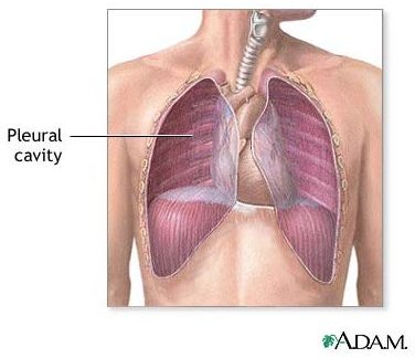
Situated between the lungs and the chest cavity, the pleural cavity is the space containing the lungs, pleura, and pleural fluid. The pleura refers to a two-layered membrane surrounding the lungs and lining the inner wall of the chest cavity. It performs to protect the lungs as well. There is a space between two layers of the pleura containing pleural fluid to lubricate and enable the lungs to move well while breathing. Click on image to enlarge.
A pleural effusion occurs when excessive fluid accumulates in the pleural cavity, then presses on the lungs, leading to difficulty breathing or shortness of breath. The lungs will not function normally at this time. Cancer can metastasize or spread to this cavity and produce excessive fluid. This condition is known as malignant pleural effusion.
Causes
A complication of cancer can lead to malignant effusion. Some cancers such as lung cancer, leukemia, breast cancer, or lymphoma might result in this condition. Difficulty breathing owing to the effusion can reduce significantly by having the fluid in the chest cavity drained.
Signs and Symptoms
Several symptoms that may occur include:
- Difficulty breathing or shortness of breath (dyspnea)
- Heavy pressing sensation on the chest
- Dry cough
- Chest pain
- Fatigue
- Fever
Diagnosis
Some tests below can help diagnose the effusion in addition to determining the exact area of it:
Thoracentesis
The doctor inserts a needle between the ribs to drain the fluid from the pleural cavity. It can help decrease compression on the lungs. In addition, a microscope can detect and analyze the fluid if cancerous cells exist.
Computed Tomography (CT) scan
CT scan makes a series of x-ray images with the aid of a computer to produce 3-D images of the internal structures and organs of the body. The doctor might inject contrast material into a vein to help increase the distinction between areas of the body.
Chest x-ray
Chest x-ray produces an x-ray image of the chest and internal organs inside the chest. The test involves radiation to the chest, resulting in a detailed image of the chest.
Biopsy
The doctor performs a biopsy unless thoracentesis works well. A biopsy is the removal of tissues, then a microscope can check whether or not cancer cells are present. The doctor takes samples by performing a thoracoscopy or surgery to examine the inner organs in the chest. He or she makes an incision between two ribs and inserts a thin and lighted tube called a thoracoscope to take samples.
Treatment
The following treatments are effective to drain the fluid from the chest:
Surgery
The doctor might perform surgery to insert a thin tube to relocate the fluid from the pleural cavity to the abdominal cavity. This procedure can help drain the fluid more certainly.
Thoracentesis
In addition to diagnosing the effusion, thoracentesis can be applied to treat it. This procedure involves a needle and/or a hollow plastic tube to collect the fluid from the pleural cavity. It will provide temporary relief from the symptoms, enable the lung to expand, and restore normal breathing.
Pleurodesis
It is a medical procedure to avoid the buildup of fluid in the space between the membranes in addition to allowing them to stick together. Once thorecentesis has removed fluid, then the doctor administers chemical agents like talc powder inside the space between the pleura. The aim of pleurodesis is to prevent the fluid from returning.
References
Robert Wood Johnson University Hospital: Malignant Pleural Effusion - https://www.rwjuh.edu/medical_services/malignant_pleural_effusion_treatment.html
The National Cancer Institute: Malignant Pleural Effusion - https://www.cancer.gov/cancertopics/pdq/supportivecare/cardiopulmonary/Patient/page3
Cancer.net: Fluid around the Lung or Malignant Pleural Effusion - https://www.cancer.net/patient/All+About+Cancer/Treating+Cancer/Managing+Side+Effects/Fluid+Around+the+Lungs+or+Malignant+Pleural+Effusion+-+ASCO+curriculum
Photo Credit
Image courtesy of the National Library of Medicine.