Jugular venous distension is a condition many are not aware of. Here we will discuss this condition and provide a detailed overview for patients.
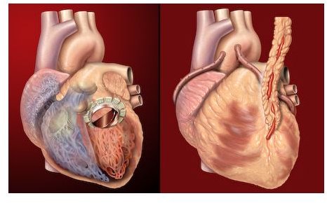
Jugular venous distention occurs as a result of high pressure in the jugular vein. When blood pressure is higher than it should be in the jugular vein, the walls of the vein can distend or swell, resulting in this condition. This condition is often linked to heart disease and cardiologists are often evaluating this vein to aid them in creating a treatment plan or making a diagnosis. The jugular vein is found on both sides of the neck. It is responsible for sending blood from the head back to the heart.
Causes
If this condition is present, meaning the patient’s jugular vein pressure is elevated, it tells his or her doctor that fluid backup from the right heart is possibly present. This may come along with tricuspid valve disease, pericardial diseases, heart failure, pulmonary stenosis, heart block, right heart malfunction, ventricular tachycardia, pulmonary hypertension, right ventricular MI, or chronic obstructive pulmonary disease (COPD).
Causes may include cardiac tamponade, hypervolemia, superior vena cava obstruction, or chronic obstructive pericarditis.
Signs and Symptoms
With jugular venous distention, the jugular vein will be visibly swollen or distended when looking at the patient’s neck. Jugular vein pressure will also be elevated.
Differential Diagnosis
During the diagnostic process, differential diagnosis is also often performed. Differential diagnosis for this condition may include:
- Congestive heart failure
- Cardiac tamponade
- Tricuspid regurgitation
- Atrial fibrillation
- Tricuspid stenosis
- Hypervolemia
- Constrictive pericarditis
- Superior vena cava syndrome
- Heart block (usually complete)
- Right ventricular infarction
- Right ventricular dilation
Diagnosis
Doctors will often begin by getting the patient’s personal medical history and performing a physical exam. The physical exam will focus on looking for normal central venous pressure, the right atrium being located five centimeters below the sternal angle, hepatojugular reflux, and no venous pulsation with compression with arterial pulsation with compression.
An ECG may show ventricular tachycardia, heart block, atrial fibrillation, right ventricular infarction, or other pathology. A chest x-ray may show cardiomegaly or congestive heart failure. An echocardiogram may be used to look for myxomas, valvular disease, or right ventricular dysfunction.
Treatment
The underlying disease will determine the treatment. Ventricular tachycardia may be treated with antiarrhythmic medications, such as lidocaine or amiodarone, ICD for recurrences, replacing electrolytes, and if hemodynamic instability is present DC countershock may be done.
A permanent pacemaker may be needed if the patient has a heart block. Tricuspid stenosis may require surgery to correct it if the patient is experiencing severe symptoms. Surgical excision may be necessary for atrial myxoma. Managing volume status is often used for constrictive pericarditis.
Resources
Family Practice Notebook. (2010). Jugular Venous Distention. Retrieved on January 10, 2010 from the Family Practice Notebook: https://www.fpnotebook.com/cv/exam/JglrVnsDstntn.htm
University of Washington Department of Medicine. (2010). Introduction: Examination of Neck Veins. Retrieved on January 10, 2010 from the University of Washington Department of Medicine: https://depts.washington.edu/physdx/neck/index.html