A rare cancer, adrenocortical carcinoma, might thrive in the adrenal cortex. Find out several tests for adrenocortical carcinoma along with their risks.
What is Adrenocortical Carcinoma?
Adrenocortical carcinoma is a cancerous tumor that grows in the adrenal cortex, which is the outer layer of the adrenal gland. Click on image to enlarge. This cancer is also known as adrenal cancer, adrenal carcinoma, or adrenocortical cancer. It is common in children below 5 years old and in adults between 30 and 40 years old. Both men and women are vulnerable to have this cancer. Genetic conditions are associated with an increased risk of this rare cancer. The cancer can generate the hormones aldosterone, estrogen, or cortisol. Common symptoms of the cancer include a lump in the abdomen, pain in the abdomen, high blood pressure, and muscle weakness.
The following are several common tests for adrenocortical carcinoma to diagnose the cancer:
Physical Exam
This kind of test can diagnose something wrong in your body including suspicious signs of cancer such as lumps or unusual things. In addition, blood tests might reveal the characteristics of adrenocortical carcinoma including increased levels of aldosterone, increased levels of cortisol, decreased levels of ACTH (adrenocorticotropic hormone), and decreased levels of potassium. Elevated aldosterone levels can lead to hypertension.
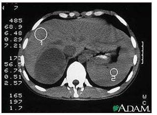
Computed Tomography (CT) Scan
A CT scan utilizes a series of x-rays taken from different angles to generate various images of tissues in the abdomen. A computer then combines all images to provide a powerful image. A CT scan might provide information about tumors including location, size, and shape. As you are exposed to radiation during this diagnosis, you might be more likely to develop cancer. A CT scan can use a dye (intravenous contrast material) to be injected into a vein to give clear images of your inner organs. However, it can result in allergic reactions including rash and hives to some people.
Position Emission Tomography (PET) Scan
A PET scan would help disclose how well your tissues in the abdomen are. A small amount of radioactive glucose is injected into a vein to diagnose tumors. The scanner travels around your body to find if there are cells taking up more glucose. Cancerous cells can come up brighter in the images because they absorb more glucose. Despite using low concentrations of radioactive glucose, this practice might endanger the fetus or a breastfeeding child. You need to consult your doctor before having this scan performed.
Magnetic Resonance Imaging (MRI) Scan
This diagnosis involves a technique applying a magnetic field, radio waves, and a computer to produce detailed images of the abdomen. After removing all metallic objects, you need to lie inside a magnetic tube, and then a large magnet aligns all the water molecules in your body. Radio waves generate signals leading to 3-D images. Doctors would notice and diagnose those images to decide further treatments. It is essential to let your doctor know if you have any metallic materials within your body, as they can distort the images. There are no dangerous side effects of a MRI scan because it is relatively safe to avoid radiation exposure.
Adrenal Angiography
This diagnosis works to examine the arteries and the blood flow near the adrenal gland. A doctor would inject a contrast material into your adrenal arteries. The doctor then takes x-rays to detect if there are blocked arteries. Allergic reactions such as rash and hives might occur due to having it performed.
References
The National Cancer Institute: General Information about Adrenocortical Carcinoma - https://www.cancer.gov/cancertopics/pdq/treatment/adrenocortical/Patient
Medline Plus: Adrenocortical Carcinoma - https://www.nlm.nih.gov/medlineplus/ency/article/001663.htm
MayoClinic.com: CT scan - https://www.mayoclinic.com/health/ct-scan/MY00309
Photo Credit
Image courtesy of the National Library of Medicine.