The cardiac nuclear stress test measures how well the heart is functioning by imaging blood flow to the heart muscle during rest and during exercise. A radioactive dye is injected into the blood stream and a scanner monitors the dye as it circulates through the heart.
What is a Cardiac Nuclear Stress Test?
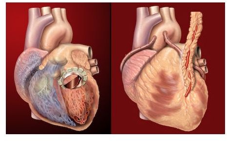
A cardiac nuclear stress test evaluates how well the heart is functioning. It images blood flow to the heart muscle during and after exercise. Areas of the heart that are damaged will have less blood flow than the rest of the heart.
The difference between a cardiac nuclear stress test and a regular exercise stress test is the imaging. This requires a radioactive dye that is injected into the bloodstream. A monitor detects the radioactivity as the dye circulates through the heart and creates an image.
Why is the Test Ordered?
A doctor uses a cardiac nuclear stress test to diagnose coronary heart disease. Usually, there are symptoms which indicate that the arteries supplying the heart are narrowing due to the accumulation of plaque. Shortness of breath and chest pain are two symptoms of coronary heart disease.
Another reason for a nuclear stress test is to determine whether the size and shape of the heart is abnormal. If a portion of the heart is enlarged, it will be visible on the imaging monitor. The test can be used to determine the ejection fraction of the heart.
Preparing for the Test
Before the test, the doctor will inform you of the preparations. Generally, you shouldn’t eat, drink or smoke within two hours of the test. It is recommended to wear comfortable clothes and walking shoes. If you have asthma, bring your inhaler and inform the medical staff of your medications.
Test Procedure
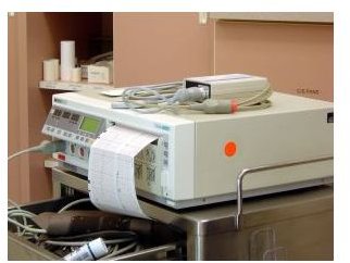
The first thing the medical staff will do is connect you to the monitoring equipment. Several electrodes will be placed on your body to monitor the heart with an electrocardiogram. A blood pressure cuff will be placed around your arm to monitor blood pressure.
The test begins at a slow walking pace and gradually ramps up in intensity. The incline of the treadmill or stationary bike will increase, which will put stress on the heart. Expect to exercise at a good pace for about 8 to 12 minutes. If you are unable to exercise, a medication is given to increase the heart rate and simulate exercise.
Once you’ve reached maximum exertion, an injection is given containing a radioactive dye. Thallium or sestamibi are two types of radioactive dyes that are introduced into the bloodstream through an IV. A monitor is used to detect the radioactivity and image the blood flow to the heart. Afterward, expect to rest between two and four hours. Then a second imaging scan is conducted to evaluate the heart when it is resting.
Results
The imaging scan will show the status of blood flow to the heart. If the blood flow is normal during rest and exercise, coronary heart disease isn’t present. If the blood flow is normal during rest, but inadequate during exercise, it may indicate a blocked artery. If the blood flow is inadequate during rest and exercise, immediate treatment for coronary heart disease is required. Parts of the heart that are not exposed to the radioactive dye contain scarred tissue caused by a heart attack.
References
1. “Nuclear Stress Test.” Mayo Clinic. https://www.mayoclinic.com/health/nuclear-stress-test/MY00994
2. “Heart Disease & Stress Tests.” WebMD. https://www.webmd.com/heart-disease/guide/stress-test--%28dupe%29